

NIH-funded researchers map basal ganglia at single-cell resolution
The resource spans human, non-human primate, marmoset and mouse data, giving scientists a common reference for neurological and psychiatric research.
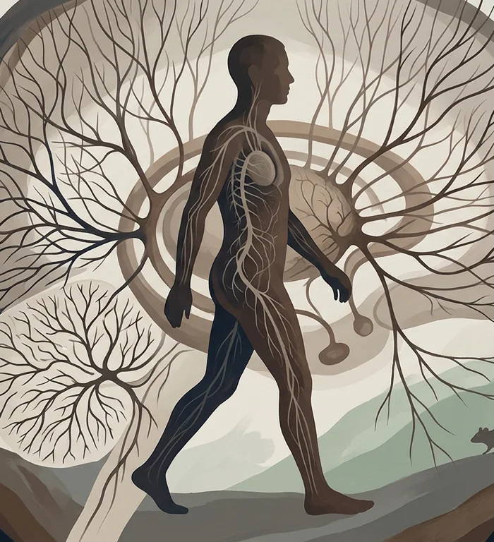
NIH-funded researchers released a 27-paper single-cell atlas of the basal ganglia on Nov. 12, mapping millions of cells across human and animal brain tissue, according to an NIH press release.
The work is basic and translational research, not a clinical test. It gives scientists a shared reference for studying how specific brain cells may contribute to neurological, psychiatric and substance-use disorders.
The atlas was produced through the NIH BRAIN Initiative and its BRAIN Initiative Cell Atlas Network. The basal ganglia are a group of brain structures involved in deliberate movement, learning and attention.
“Representing the brain at this level of granularity opens so many opportunities to better understand basic brain function and the cellular underpinnings of devastating neurologic, neuropsychiatric, and substance use disorders,” John Ngai, director of the NIH BRAIN Initiative, said in the press release.
Researchers used single-cell sequencing, a method that reads genetic activity in individual cells, on donated human brain tissue after death. They then linked those cell features to three-dimensional locations within the basal ganglia.
The collection includes data from mature and developing human brain tissue, as well as non-human primate, marmoset and mouse brain data. NIH said the multi-species design is intended to support work that connects human biology with preclinical models.
One autism-related paper used the atlas to organize sequencing data from humans who had autism, according to NIH. That work pointed to possible disruptions in cell populations in the striatum, a major input region of the basal ganglia, and suggested deeper brain circuits may be involved alongside cortical mechanisms.
“These multi-species atlases have unlocked important new findings about how the brain develops and functions at the single-cell level,” Amy Bany Adams, acting director of the National Institute of Neurological Disorders and Stroke, said in the release.
The collection also includes a mouse-model Parkinson’s disease paper. NIH said scientists used an enhancer, a genetic control element, to increase a dopamine precursor in striatum cells, easing symptoms while reducing the severity of common side effects in mice.
The study cannot show that the atlas can diagnose autism, explain autism causes in individuals or treat Parkinson’s disease in people. The Parkinson’s finding is from mouse models, and the autism finding is framed by NIH as a potential cell-population disruption.
The infrastructure may matter as much as the atlas itself. NIH said BICAN-funded scientists created open-source tools, including preprocessing software, a web platform for exploring basal ganglia data and the Neuroanatomy-anchored Information Management Platform, or NIMP, to align donated human samples within a common 3D brain reference.
“The findings presented in these studies are scientifically profound in and of themselves. But what is perhaps just as notable is how much computational infrastructure these researchers have created to support and shape the future of neuroscience,” Andrea Beckel-Mitchener, acting director of the National Institute of Mental Health, said in the release.
The 27 papers appear in Cell, Cell Reports Medicine, Cell Systems, Cell Genomics and Neuron, according to NIH. The agency said it supported the work through the BRAIN Initiative, co-led by the National Institute of Mental Health and the National Institute of Neurological Disorders and Stroke.
Source: NIH press release, Nov. 12, 2026.
Atlas360 reports health research and does not provide medical advice.
More stories
- PFOS levels track 93% higher aggressive breast cancer rate
- Fructose may weaken ovarian cancer cell bonds after chemotherapy
- High-intensity sports raise bone markers by up to 46%
- Plant-based dairy swaps may lower iodine intake
- Adults 50 to 64 reach 7.5% heavy-drinking rate
- Dinch exposure correlates with 27-gram lower birth weight
Latest news
- FAA halts US departures after communications disruption
- Europe faces unity test as Trump revives Ukraine diplomacy
- Democrats push AI regulation as two state veterans arrive
- Lutnick faces AI chip scrutiny as Bessent faults Commerce
- AI investment boom binds global suppliers to US spending
- Senate Democrats face leadership fight after midterm gains
- Trump extends Jones Act waiver for another 90 days: official
- Russia reports 1% growth as refinery output declines 18%