

Scientists test human regenerative powers via growth factors
A Texas A&M-led study reports a two-step growth-factor approach that shifted mammalian wound repair away from scarring and toward partial digit tissue…
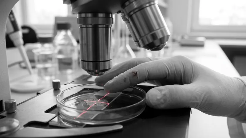
Human regenerative powers may be less absent than dormant, according to Texas A&M researchers who report a method that nudged mammalian healing toward regrowth instead of scarring.
In work published June 17, 2026, in Nature Communications , the team described a two-stage, growth-factor treatment that restored multiple digit structures after amputation in animal studies. The regrown anatomy included bone and joint components, along with connective tissues such as ligaments and tendons.
The findings challenge a long-running assumption that mammals fundamentally lack the capacity to rebuild complex parts once they are lost. Instead, the study frames regeneration as a program that may be present but typically overridden by the body’s rapid wound-closing response.
A two-stage treatment aims to shift the healing program
Mammals usually respond to severe injury by sealing tissue quickly through fibrosis, a process driven largely by fibroblast cells. That reaction is protective because it reduces exposure and infection risk, but it often produces scar tissue that can block the organized reconstruction needed to restore original form and function.
By contrast, classic regenerators such as salamanders rely on cells that assemble into a “blastema,” a temporary, growth-focused structure that supports rebuilding. Texas A&M’s study focused on whether mammalian fibroblasts at the injury site could be steered away from scar-building behavior and toward a blastema-like path.
The researchers tested a sequential approach using two established growth factors: fibroblast growth factor 2 (FGF2) and bone morphogenetic protein 2 (BMP2). The sequence, timing, and intent were designed to intervene after the body had already executed its initial wound-closure steps, rather than trying to suppress them from the start.
According to the report, the first stage involved applying FGF2 after the wound surface had closed. The goal was to redirect the local repair environment toward a regenerative state rather than a purely fibrotic one.
The second stage introduced BMP2, a signaling molecule known for its role in bone formation and skeletal development. In the animal experiments described, this paired strategy supported the return of several digit elements following amputation, including skeletal and joint-related structures.
What the study says about scars, fibroblasts, and regrowth
At the center of the work is the idea that fibroblasts can follow divergent outcomes after injury. In typical mammalian repair, fibroblasts help create a collagen-rich matrix that closes and stabilizes the wound but can leave stiff, function-limiting scar tissue.
The Texas A&M group argues the same broad cell population can be prompted to support a different architecture—one more compatible with organized tissue rebuilding. In that framing, the critical barrier is not the absence of regenerative cells, but a default biological “decision” that prioritizes rapid sealing over reconstruction.
Ken Muneoka, a professor at Texas A&M’s College of Veterinary Medicine and Biomedical Sciences, said the question of why some animals regenerate and humans do not has persisted since ancient times. The new study, he and colleagues suggest, provides experimental evidence that mammalian repair pathways can be redirected under specific conditions.
The team also emphasized that the regrown tissues were not exact replicas of the originals. Even so, the ability to regain multiple integrated structures—bone, joint components, and connective tissues—in a coordinated way is a notable step beyond approaches that only accelerate closure or reduce inflammation.
Implications for amputations and tissue repair research
The work arrives amid broader interest in improving outcomes after amputations and complex injuries, where scarring can limit mobility, contribute to chronic pain, and complicate prosthetic fit. A method that reduces fibrosis while encouraging structured regrowth could, if translated, change how clinicians think about post-injury recovery.
However, the report is based on animal studies, and the pathway to human use would require extensive replication, safety profiling, and careful testing of dosing and timing. Growth factors such as BMP2 have known biological potency, making delivery methods and side-effect management central considerations for any future clinical development.
Next steps for the research field will likely include identifying which mammalian injuries and tissues respond best to this kind of sequential signaling, and determining how closely regenerated structures can match original anatomy and function. For now, the study’s main contribution is a clearer experimental roadmap for exploring whether a latent regenerative program can be switched on in mammals.
More stories
- Deadlock’s Solomon hides 8-to-32 chess reload boards
- Nintendo Life polls 673 readers on Ocarina pronunciation
- Bandai Namco says Ace Combat 8 sells over 1 million copies
- Kingdom Hearts holds No. 1 on Switch 2 eShop chart
- Cyberpunk 2077 tops 40m sales as Paramount develops film, Odatv says
- L3Harris says 60 Wolf Pack flights tighten defense buying options
- Government curbs on AI models add uncertainty for investors
- NASA taps Relativity Space to deliver Aeolus Mars mission
- Microsoft Defender Misflags DigiCert Root Certificates, Triggering Windows Trust Store Disruptions
- 007 First Light's main campaign runs about 20 hours
Latest news
- Merck KGaA tops MediciNova on 8 of 15 metrics
- Tanker burns near Hormuz after munition strike
- Donor-specific genomes reveal 1,790 chromatin hotspots
- Trump claims Ukraine-Russia energy ceasefire agreement
- Zelensky condemns US-Russia diesel deal as Putin gift
- Trump announces Russia-Ukraine energy ceasefire as fuel prices climb
- HCW Biologics leads Cardio Diagnostics on 9 of 14 measures
- Lebanon seizes 500 kg explosives tied by officials to Hezbollah